Research Projects
- Gut and stomach metamorphosis in the common clownfish
- Evolution of the bilaterian throughgut
- Endocrine disrupting effects of organophosphate pesticides
- Effect of dietary phytochemicals on coral reef biology
- Endoderm development in stem-cell-based models of early mouse development
Postdoctoral research PhD research
Gut metamorphosis in the common clownfish
I study the hormonal control of metamorphosis in the common clownfish (Amphiprion ocellaris). Specifically, I focus on the transformations in gastrointestinal anatomy and function as the larvae undergo the broader metamorphic changes that prepare them to settle in the reef.
Work related to this project is ongoing, though below are related publications:
Roux, Vianello, Laudet; "Physiology of metamorphosis", 2023;
Zwahlen, Gairin, Vianello, Mercader, Roux, Laudet; "The ecological function of thyroid hormones", 2024.

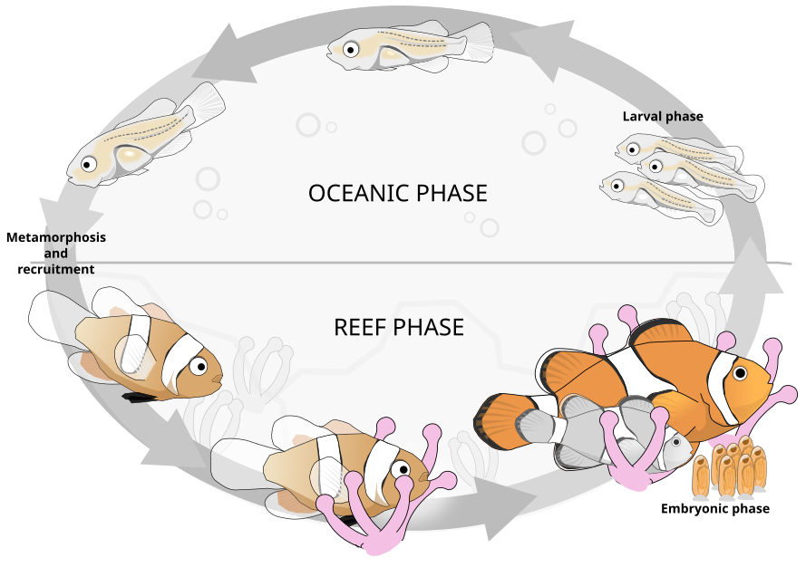
Illustration of the lifecycle of the common clownfish. Larvae (rightmost) disperse into the ocean (pelagic phase). Major physiological, behavioral, and ecological changes (metamorphosis) take place as the larval body plan is recruited into the reef and the clownfish finds an anemome in which to house. Figure redrawn from (Roux et al. 2019).  Image licensed under a Creative Commons Attribution-NonCommercial-ShareAlike 4.0 International License.
Work related to this project is ongoing, though below are related publications:
Roux, Vianello, Laudet; "Physiology of metamorphosis", 2023;
Zwahlen, Gairin, Vianello, Mercader, Roux, Laudet; "The ecological function of thyroid hormones", 2024.

 Image licensed under a Creative Commons Attribution-NonCommercial-ShareAlike 4.0 International License.
 EcoEvoDevo endoderm metamorphosis gut development
Evolution of the bilaterian throughgut
An EcoDevo exploration of gut organisation across marine bilaterians
My main postdoctoral research project, a collaboration with three other Academia Sinica labs to bring together the study of a total of five different marine organisms on the common topic of gastrointestinal development and throughgut evolution.
We considered five marine organisms across main branches of the bilaterian evolutionary tree. As a representative of the protostomes, the annelid worm Platynereis dumerilii (Dumeril's clam worm; Audouin & Milne Edwards, 1833). Among deuterostomes, within the Ambulacraria, the echinoderm Strongylocentrotus purpuratus (purple sea urchin; Stimpson, 1857) and the hemichordate Ptychodera flava (acorn worm; Eschscholtz, 1825). For the chordate branch, the cephalochordate Branchiostoma floridae (Florida lancelet; Hubbs, 1922), and the vertebrate Amphiprion ocellaris (common clownfish; Cuvier, 1830). For each We develop spatiotemporal transcriptomic resources along the entire gut, in the adults, the larval stages, amd across the metamorphic process.
One first manuscript our of this collaboration has now been published:
Vianello, Lin, Pinem, Li, Li, Sonia, Lee, Wu, Laudet, Su, Yu, Schneider: "Deconstructing the common anteroposterior organisation of adult bilaterian guts"

The annelid worm Platynereis dumerilii, the echinoderm Strongylocentrotus purpuratus, an acorn worm, a cephalochordate Branchiostoma, and the vertebrate Amphiprion ocellaris [Image sources: Torkild Bakken; Flickr/kqedquest; Chris Lowe; D. L. GEIGER/SNAP/ALAMY; Unsplash]
We considered five marine organisms across main branches of the bilaterian evolutionary tree. As a representative of the protostomes, the annelid worm Platynereis dumerilii (Dumeril's clam worm; Audouin & Milne Edwards, 1833). Among deuterostomes, within the Ambulacraria, the echinoderm Strongylocentrotus purpuratus (purple sea urchin; Stimpson, 1857) and the hemichordate Ptychodera flava (acorn worm; Eschscholtz, 1825). For the chordate branch, the cephalochordate Branchiostoma floridae (Florida lancelet; Hubbs, 1922), and the vertebrate Amphiprion ocellaris (common clownfish; Cuvier, 1830). For each We develop spatiotemporal transcriptomic resources along the entire gut, in the adults, the larval stages, amd across the metamorphic process.
One first manuscript our of this collaboration has now been published:
Vianello, Lin, Pinem, Li, Li, Sonia, Lee, Wu, Laudet, Su, Yu, Schneider: "Deconstructing the common anteroposterior organisation of adult bilaterian guts"

EvoDevo throughgut Anteroposterior patterning
Endocrine disrupting effects of organophosphate pesticides
using clownfish as a laboratory model
Read the preprint at: Reynaud, Vianello et al., "The multi-level effect of chlorpyrifos during clownfish metamorphosis" (2024).
A collaboration with Dr. Matthieu Reynaud. [Pharmacological perturbation, transcriptomics, fish phisiology]. The collaboration is still ongoing with more data and an extension of the ecotoxicological relevance of the study.
Chlorpyrifos is an organophosphate pesticide widely used in agriculture: classified as a persitant organic polluter, it leaches into the soil and waterstreams following application, and accumulates long-term within coastal marine sediments. Its use has bee long associated with the violation of labour- and human rights in both Southeast Asia and South America, backed at least in part by neocolonial EU trade policies. In Taiwan, the use of Chlorpyrifos (陶斯松) is under ongoing phaseout, with a total ban planned by 2026 . Within South East Asia, its use has been officially banned in Malaysia, Vietnam, and Thailand, though not yet in the Philippines and Indonesia, despite this pesticide having such "significant adverse human health and/or environmental effects, that global action is warranted" according to the latest Stockholm Convention's Persistent Organic Pollutants Review Committee review .
Particular alarm and morbidity has always concerned the effects of Chlorpyrifos as an endocrine disruptor, including on the Thyroid Hormone Axis. By using metamorphosing larvae of the common clownfish Amphiprion ocellaris as a model system of Thyroid Hormone endocrine regulation in fish, we characterised the effect of this pesticide on a marine fish at the level of the whole transcriptome, with a further focus on the consequences on the Thyroid Hormone endocrine axis. Beside the ecotoxicological inferences that this research allows, specifically on the threat represented by transmaritime trade across tropical waters, our findings provide one of the first system-level description of the potential effects of this pesticide on fish, and in comparison to known thyroid hormone axis modulators.

Chlorpyrifos pesticide spraying over agriculture crops
A collaboration with Dr. Matthieu Reynaud. [Pharmacological perturbation, transcriptomics, fish phisiology]. The collaboration is still ongoing with more data and an extension of the ecotoxicological relevance of the study.
Chlorpyrifos is an organophosphate pesticide widely used in agriculture: classified as a persitant organic polluter, it leaches into the soil and waterstreams following application, and accumulates long-term within coastal marine sediments. Its use has bee long associated with the violation of labour- and human rights in both Southeast Asia and South America, backed at least in part by neocolonial EU trade policies. In Taiwan, the use of Chlorpyrifos (陶斯松) is under ongoing phaseout, with a total ban planned by 2026 . Within South East Asia, its use has been officially banned in Malaysia, Vietnam, and Thailand, though not yet in the Philippines and Indonesia, despite this pesticide having such "significant adverse human health and/or environmental effects, that global action is warranted" according to the latest Stockholm Convention's Persistent Organic Pollutants Review Committee review .
Particular alarm and morbidity has always concerned the effects of Chlorpyrifos as an endocrine disruptor, including on the Thyroid Hormone Axis. By using metamorphosing larvae of the common clownfish Amphiprion ocellaris as a model system of Thyroid Hormone endocrine regulation in fish, we characterised the effect of this pesticide on a marine fish at the level of the whole transcriptome, with a further focus on the consequences on the Thyroid Hormone endocrine axis. Beside the ecotoxicological inferences that this research allows, specifically on the threat represented by transmaritime trade across tropical waters, our findings provide one of the first system-level description of the potential effects of this pesticide on fish, and in comparison to known thyroid hormone axis modulators.

EcoDevo Pesticides metamorphosis Endocrine disruption
Effect of dietary phytochemicals on coral reef biology
The bisindolic alkaloid Caulerpin and its effects on fish metabolism and physiology
A collaboration with Carmela Celentano, Italy. The results from this research have not yet been published.
Species of the genus Caulerpa, a siphonaceous green alga that grows in the upper sublittoral zone of tropical coral reefs, are widely distributed in Australian and Southeast Asian waters. Caulerpa lentillifera and varieties of Caulerpa racemosa are for example considered a delicacy in the Philippines, Indonesia, Malaysia, and Okinawa (Japan), and many Caulerpa species have (also) been long valued for their medicinal uses. In fact, Caulerpa varieties are now widely farmed in many Southeast Asian nations, a practice first developed in the 1950s in Mactan Island (Cebu, Philippines).
On the other hand, a member of the Caulerpa racemosa complex is currently a source of heightened concern in the Mediterranean Sea, new waters it is believed to have reached as a result of aquarium trade from Western Australia. In these waters, in the form of the so-called var. cylindracea, this species has shown one of the highest invasive characters ever recorded in the Mediterranean, affecting pre-established seagrass ecosystems and reducing species diversity. Further concern has notably derived from its accumulation within the tissues of local sparid species of human consumption (e.g. Diplodus sargus).
Critically, Caulerpa cylindracea and many other species of the genus synthesize a bisindolic red pigment called caulerpin, first discovered in Caulerpa racemosa by Filipino scientist Gertrudes Aguilar Santos. While data on the accumulation of this compound in fish of historical Caulerpa spp. tropical habitats are difficult to find, an increasing body of literature has instead documented its clear accumulation in muscle and liver of Mediterranean species. The effects of caulerpin on fish physiology and metabolism have however been only characterised in terms of a restricted panels of expected targets or target processes (notably, oxidative stress, selected lipid metabolism enzymes, etc...). By leveraging the amenability of Amphiorion clarkii to experimentation, our collaborative research characterised the effects of caulerpin at all levels of fish physiology: transcriptomics, lipidomics, metatranscriptomics, behaviour .
A field of Caulerpa spp. in the waters around Bantayan Island (Cebu, Philippines). From Rey Baruc's video "Lato or Sea Grapes ( Green Caviar ) in Bantayan Cebu"
Species of the genus Caulerpa, a siphonaceous green alga that grows in the upper sublittoral zone of tropical coral reefs, are widely distributed in Australian and Southeast Asian waters. Caulerpa lentillifera and varieties of Caulerpa racemosa are for example considered a delicacy in the Philippines, Indonesia, Malaysia, and Okinawa (Japan), and many Caulerpa species have (also) been long valued for their medicinal uses. In fact, Caulerpa varieties are now widely farmed in many Southeast Asian nations, a practice first developed in the 1950s in Mactan Island (Cebu, Philippines).
On the other hand, a member of the Caulerpa racemosa complex is currently a source of heightened concern in the Mediterranean Sea, new waters it is believed to have reached as a result of aquarium trade from Western Australia. In these waters, in the form of the so-called var. cylindracea, this species has shown one of the highest invasive characters ever recorded in the Mediterranean, affecting pre-established seagrass ecosystems and reducing species diversity. Further concern has notably derived from its accumulation within the tissues of local sparid species of human consumption (e.g. Diplodus sargus).
Critically, Caulerpa cylindracea and many other species of the genus synthesize a bisindolic red pigment called caulerpin, first discovered in Caulerpa racemosa by Filipino scientist Gertrudes Aguilar Santos. While data on the accumulation of this compound in fish of historical Caulerpa spp. tropical habitats are difficult to find, an increasing body of literature has instead documented its clear accumulation in muscle and liver of Mediterranean species. The effects of caulerpin on fish physiology and metabolism have however been only characterised in terms of a restricted panels of expected targets or target processes (notably, oxidative stress, selected lipid metabolism enzymes, etc...). By leveraging the amenability of Amphiorion clarkii to experimentation, our collaborative research characterised the effects of caulerpin at all levels of fish physiology: transcriptomics, lipidomics, metatranscriptomics, behaviour .

Ecology and Physiology Caulerpin Invasive species Phytochemicals
Endoderm development in gastruloids
In humans, mice, and other mammals key internal organs such as the gut, lungs, pancreas, liver, thyroid, thymus, and bladder (and many others!) all derive from the same embryonic tissue: the endoderm. The development of all of these structures thus depends on a same set of early cells, and on the developmental instructions provided to them by the embryo. To study what these instructions might be, I am currently investigating the modes of development that endoderm cells (and their progenitors) execute when they can not rely on them. That is, when they are made to self-organise in vitro in a dish in the lab. Even on their own, endoderm cells emerge with timings and patterns comparable to what happens when they are on the embryo, and eventually form patterned structures in many way similar to the embryonic gut tube. My research touches on topics of endoderm and epithelial identity, self-organisation, patterning, and fundamental mammalian developmental biology.

Embryonic stem cells can be left to self-organise into structures called Gastruloids. These provide an in vitro model of embryonic development. Endoderm cells, marked here in cyan (FoxA2), form an elongated internal core. The posterior marker TBra (gold) marks the posterior of the aggregate.

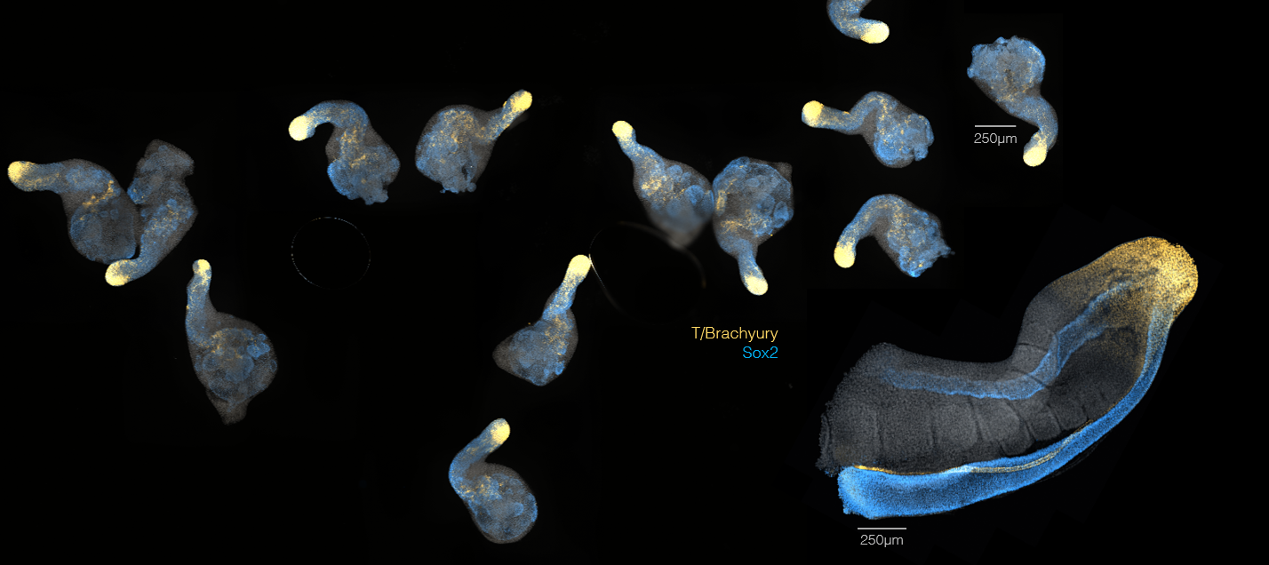
Gastruloids have been found to express genes in a organised way, in many way similar (and in many ways different) from that of the real mouse embryo. In the image above, the same markers have been stained in the tail of a mouse embryo (big structure on the right) and in 7-days-old Gastruloids. Insight comes from the comparison between what seen in the embryo and what observed to happen in these self-organising *in vitro* systems.

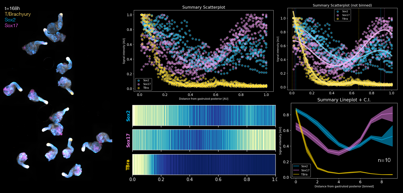
The patterns of gene expression amongst Gastruloids are very robust and reproducible. We can image entire populations of Gastruloids and extract spatial information about where specific genes are expressed along the main axis.


gastruloids endoderm embryonic stem cells
